https://goo.gl/maps/uaT85k2pLJNKD4Jp8
TELEGRAM
Γιά Ελληνικά προχωρείστε προς τα κάτω
The patient is hospitalized at the Athens Medical Center
by the medical team of the Oncology Department
under the guidance of
Oncologist
Professor K. Syrigos
The doctors of the team
Ilias Karabelas Pathologist
Niki Marioli Specialist physician
Sophia Tsagouli Pulmonologist
Vasilis Ramfidis Pathologist-Oncologist
Despoina Nasi Oncologist
Associate pulmonologist
Alexandros Viskos
Head of one day hospitalization department
Panagiota Balomenou

Η ασθενής νοσηλεύεται στο Ιατρικό Κέντρο Αθηνών
από την ιατρική ομάδα του Ογκολογικού Τμήματος, υπό την καθοδήγηση του
Ογκολόγου
Καθηγητού κ. Κ. Συρίγου
Οι Γιατροί της ομάδας
Σοφία Τσαγκούλη Πνευμονολόγος
Βασίλειος Ραμφίδης Παθολόγος-Ογκολόγος
Δέσποινα Νάση Ογκολόγος
Συνεργαζόμενος ιατρός πνευμονολόγος
Αλέξανδρος Βίσκος
Επικεφαλής Ημερίσειας Νοσηλείας
Παναγιώτα Μπαλωμένου
===================================================================
FOLFIRINOX TREATMENT DETAILS
First 3-months, 6 therapies FOLFIRINOX cycle
FOLFIRINOX DOSES
IRITEC 2fl 100mg/5 ml Dose: 200mg campto
OXAVIATIN 1fl 100mg/20ml Dose: 100mg
LEUCOVORIN 3fl 200 mg/20 ml vial Dose: 550mg
5 FLUOROURACIL (5FU) 1fl 5g
Dose 5FU bolus: 550 mg
Dose 5FU in 48 hours: 2400 mg
Initial value of CA 19-9: 273 (maximum normal value: 39)
CT scan DIAGNOSIS
However treatment continued, without any action to correct this catheter dysfunction.
Στη θεραπεία αυτή σταμάτησε να υπάρχει επιστροφή αίματος με την αναρρόφηση, μέσω του φλεβοκαθετήρα του portacath. Αυτή η δυσλειτουργία συνεχίστηκε σε όλες τις ακόλουθες θεραπείες αυτού του κύκλου. Ταυτόχρονα, υπήρχε έντονος ερεθισμός του δέρματος στο σημείο εισόδου του καθετήρα μέσα στη φλέβα και έντονος συνεχής πόνος στην ωμοπλάτη της ασθενούς, χωρίς καμία ορθοπεδική αιτία, όπως 'εδειξαν οι μαγνητικές τομογραφίες που έγιναν.
Κατόπιν τούτου υπάρχει το ενδεχόμενο ποσότητα του 5FU της 48ωρης χορήγησης, να διέφευγε προς στους ιστούς από το σημείο που ο καθετήρας εισήρχετο στην βραχιονοκεφαλική φλέβα και είχε ερεθιστεί έντονα όλη η περιοχή και να μην εισήρχετο στην κυκλοφορία του αίματος.
Ωστόσο, η θεραπεία συνεχίστηκε χωρίς καμία ενέργεια για τη διόρθωση αυτής της δυσλειτουργίας του καθετήρα.
3rd therapy: June 12, 2019 CA 19-9 after 12 days:22,1
4th therapy: June 26, 2019 CA 19-9 after 12 days:21,3
5th therapy: July 10, 2019 CA 19-9 after 12 days:19,1
6th therapy: July 24, 2019 CA 19-9 after 12 days:29,8
End of 2nd FOLFIRINOX CYCLE therapy
CT scan DIAGNOSIS
Χωρίς αξιόλογη μεταβολή των προύπαρχουσών οριακών λεμφαδένων της ρίζας του μεσεντερίου με συρρέουσα μορφολογία.


FOLFIRINOX DOSES
IRITEC 2fl 100mg/5 ml Dose: 150mg campto
OXAVIATIN 1fl 100mg/20ml Dose: 80mg
LEUCOVORIN 3fl 200 mg/20 ml vial Dose: 550mg
5 FLUOROURACIL (5FU) 1fl 5g
No 5FU bolus dose
Dose 5FU in 48 hours: 2550 mg
Anti-thrombotic treatment: INNOHEP (TINZAPARIN SODIUM) INJ.SOL 14000anti-XalU/0,7ML PF.SYR BTx10PF.SYRSx0,7ML injections. Daily dose was set at 0,55ML
Initial value of CA 19-9: 49 (maximum normal value: 39)
1st therapy: September 30, 2019 CA 19-9 after 12 days: 80,1
2nd therapy: October 15, 2019 CA 19-9 after 12 days: 81,4
3rd therapy: October 29, 2019 CA 19-9 after 12 days:112
4th therapy: November 13, 2019 CA 19-9 after 12 days:112
Imaging scans details - Λεπτομέρειες απεικονιστικών
Click on the following texts to get them sharper
Κάντε κλικ επάνω στα παρακάτω κείμενα για να δείτε με ευκρίνεια

===================================================================
το εντέρο είναι φυσιολογικό. Μετά ταύτα, επειδή το πάγκρεας χαρακτηρίζετο ως φυσιολογικό στην προηγούμενη μαγνητική τομογραφία της 4ης Μαίου 2018, ο θεράπων ιατρός συνέστησε επανάληψη της ανωτέρω μαγνητικής τομογραφίας μετά τρίμηνο, πράγμα το οποίο και έγινε


Πατείστε στην εικόνα για να μεγαλώσει

===================================================
The patience started in October 4th, 2018, a 3 months chemotherapy with
ABRAXANE (nab-paclitaxel)
and GEMNIL (gemcitabine)
Η ασθενής άρχισε στις 4/10/2018 τρίμηνη χημειοθεραπεία με ABRAXANE (nab-paclitaxel)
και GEMNIL (gemcitabine)
===================================================
January 7, 2019
UPPER AND LOWER ABDOMEN CT
Upper and Lower and Chest CT of January 7, 2019, following the end of the first 3 months cycle of chemotherapy
Download and Review the CT
One Drive page
Αξονική Τομογραφία της 7ης Ιανουαρίου 2019, μετά το πέρας
του πρώτου τρίμηνου κύκλου της χημειοθεραπείας
Download and Review the CT
Κατεβάστε και Δείτε την αξονική τομογραφία
One Drive page
TELEGRAM
Γιά Ελληνικά προχωρείστε προς τα κάτω
Medical report of the patient F. L. from Athens Greece,
with liver metastatic pancreatic cancer
Age: 78
Performance Status: 0
Height: 1,58 m.
Weight: 47 kg (from 58 kg in April 2018, before starting the cancer problem)
The patient started having problems with digestion and anorexia in April 2018. After many medical tests she had, in September 2018 was diagnosed that in fact, there was a 4.8 cm. metastatic to liver, tumor in the uncinate process of the pancreas. So the patience started in October 4th, 2018, a 3 months chemotherapy with ABRAXANE (nab-paclitaxel) and GEMNIL (gemcitabine), but the results were very poor. CT scan showed somewhat larger dimensions of the tumor in the pancreas and secondary liver lesions in most parts of the liver, with the exception of pre-existing lesions in section VI, which were mostly receding. Carbohydrate antigen 19-9 (CA 19-9) from 114 which was before starting the treatment, reached to 273 after the end of it.. All other organs, including lungs were normal.
So in January 21, 2019, the patient started a new 3 months (twice a month) cycle of chemotherapy with FOLFIRINOX (modified): Campto (Irinotecam), Eloxatin (Oxaliplatin), Leucovorin, 5FU.
This cycle ended in April 2019 and it seems that the patient benefits from it, as the cancer marker CA 10-9, has fallen from 387 to 45 units of value (max normal value being 39 units) and according to the CT scan, there is reduction per size of the primary lesion at pancreas (almost 50% shrinking of the tumor), as well as of secondary localizations at liver. But there is a small increase of the pleural effusion volume at pelvis minor and slightly increased size and confluent morphology of the pre-existing marginal lymph nodes at the mesenteric root.
So the patient started having the same therapy for another 3-months cycle. This cycle ended at July 24 2019 and at August 23 2019 the patient had CT scan which showed the following findings:
Upper and Lower abdomen CT Minor degree increase of size of the primary lesion at pancreas which infiltrates the upper mesenteric artery which is made indiscernible at minor extend (novel finding). Minor degree reduction of the secondary localization at liver while another one is depicted without notable differentiation. Two other secondary localizations are depicted with differentiation of their morphology presenting hypodense center at present study versus areas of increased contrast enhancement at prior study. The pleural fluid effusion at pelvis minor is not depicted at prior study while novel finding consist the few small intestine helices with thickened wall. Without notable differentiation of the pre-existing marginal lymph nodes at the mesenteric root with confluent morphology.
Chest CT Findings suspicious of secondary localizations not detected also at present study. Novel finding consists the small pleural fluid effusion at left as well as the atelectatic sings at the left pulmonary base and at the apical segment of the lower left lobe. Novel finding consists also the absence of contrast enhancement at the left brachiocephalic vein alongside the catheter’s course, a finding suspicious of thrombosis, further study with computed venography is recommended.
Finally, the suspected thrombosis of the brachiocephalic vein at the catheter course, was confirmed by computed venography. Thrombosis which, in combination with the severe irritation present at the catheter entry site into the vein and
the severe pain in the left shoulder blade, led to the decision to remove the portacath from the left side of the chest and to place a new one, at the right side of the chest and in the jugular
vein. Thereafter it was decided to
continue the FOLFIRINOX treatment for 3 more months, which began on September 30,
2019.
The patient passed away in January 2nd 2020 of ileum!
The patient is hospitalized at the Athens Medical Center
by the medical team of the Oncology Department
under the guidance of
Oncologist
Professor K. Syrigos
The doctors of the team
Ilias Karabelas Pathologist
Niki Marioli Specialist physician
Sophia Tsagouli Pulmonologist
Vasilis Ramfidis Pathologist-Oncologist
Despoina Nasi Oncologist
Associate pulmonologist
Alexandros Viskos
Head of one day hospitalization department
Panagiota Balomenou
Ιατρικό ιστορικό της ασθενούς Φ. Λ.
με ηπατικό μεταστατικό καρκίνο παγκρέατος
με ηπατικό μεταστατικό καρκίνο παγκρέατος
Ηλικία: 78 ετών
Performance Status: 0
Υψος: 1,58 μ.
Βάρος: 47 κιλά (από 58 κιλά που ήταν τον Απρίλιο του 2018, πριν να αρχίσει το πρόβλημα)
Η ασθενής άρχισε να έχει προβλήματα δυσπεψίας και μειωμένη όρεξη τον Απρίλιο του 2018. Εκανε γαστροσκόπηση η οποία ήταν αρνητική για ευρήματα και τον Μάϊο του 2018 μαγνητική τομογραφία άνω - κάτω κοιλίας, η οποία έδειξε ότι όλα τα όργανα ήταν φυσιολογικά πλην όμως υπήρχε "πάχυνση του λεπτού εντέρου ισοϋψώς των προσθίων-κατωτέρω ορίων του παγκρέατος" (διάγνωση η οποία ήταν λανθασμένη όπως απεδείχθη εκ των υστέρων..). Κατόπιν αυτού ο θεράπων ιατρός, επειδή στην διάγνωση αυτή ανεφέρετο (λανθασμένα) ότι το πάγκρεας είναι φυσιολογικό και μετά από εντεροσκόπηση που έκανε χωρίς ευρήματα πάχυνσης του εντέρου, συνέστησε επανάληψη της μαγνητικής τομογραφίας τον Σεπτέμβριο του 2018, στην οποία απεκαλύφθη ότι στην πραγματικότητα υπήρχε όγκος στην αγκιστροειδή απόφυση του παγκρέατος, ο οποίος είχε διπλασιαστεί από τον Μάϊο μέχρι τον Σεπτέμβριο (από 3,5 εκ. έγινε 4,8 εκ.) και είχε κάνει και πολλαπλές μεταστάσεις στο ήπαρ..
Κατόπιν αυτού η ασθενής άρχισε χημειοθεραπεία στις 4 Οκτωβρίου 2018 διάρκειας 3 μηνών με ABRAXANE (nab-paclitaxel) και GEMNIL (gemcitabine) πλην όμως, όπως απεδείχθη από αξονική τομογραφία που έγινε στις 7 Ιανουαρίου 2019, τα αποτελέσματα ήταν πολύ πτωχά έως ανύπαρκτα, με επιδείνωση της κατάστασης, ως κατωτέρω:
"Λίγο μεγαλύτερες διαστάσεις εμφανίζει προϋπάρχουσα εξεργασία στην αγκιστροειδή απόφυση του παγκρέατος. Δευτεροπαθείς εντοπίσεις ήπατος στα περισσότερα τμήματα του ήπατος με εξαίρεση τις προϋπάρχουσες αλλοιώσεις στο τμήμα VI κυρίως, οι οποίες εμφανίζουν υποχώρηση"
Ολα τα άλλα όργανα συμπεριλαμβανομένων των πνευμόνων ήταν φυσιολογικά.
Επίσης η τιμή του CA 19-9 από 114 που ήταν προ της ενάρξεως της ανωτέρω χημειοθεραπείας, έφθασε στα 273 μετά το πέρας της.
Κατόπιν αυτού η ασθενής άρχισε στις 21 Ιανουαρίου 2018, νέο κύκλο τρίμηνης χημειοθεραπείας (με δόσεις 2 φορές τον μήνα) με FOLFIRINOX (modified): Campto (Irinotecam), Eloxatin (Oxaliplatin), Leucovorin και 5FU. Μετά το πέρας του κύκλου της θεραπείας αυτής διαπιστώθηκε ότι η ασθενής ανταποκρίθηκε θετικά. Συγκεκριμένα ο καρκινικός δείκτης έπεσε από το 397 στο 45 και σύμφωνα με το συμπέρασμα της αξονικής τομογραφίας που έγινε, υπάρχει ελάττωση των διαστάσεων της πρωτοπαθούς εξεργασίας (σμίκρυνση του όγκου κατά 50% περίπου), καθώς επίσης και των δευτεροπαθών εντοπίσεων του ήπατος. Παρατηρείται ελαφρώς αυξημένο μέγεθος και συρρέουσα μορφολογία προϋπαρχόντων οριακών λεμφαδένων της ρίζας του μεσεντερίου.
Κατόπιν τούτων απεφασίσθη η συνέχιση της ιδίας θεραπείας για άλλο 1 τρίμηνο.
Μετά το πέρας και του δεύτερου τριμήνου χημειοθεραπείας με Folfirinox, καθ'όλη τη διάρκεια του οποίου ο καρκινικός δείκτης CA 19-9 ήταν φυσιολογικός (με τιμές από 19 μέχρι 32, με ανώτατο φυσιολογικό όριο το 39), η ασθενής παρουσίασε:
Μικρού βαθμού αύξηση των διαστάσεων της πρωτοπαθούς εξεργασίας του παγκρέατος (από 41 χ 24χιλ. έγινε 47 χ 26χιλ.)η οποία διηθεί την άνω μεσεντέριο αρτηρία η οποία καθίσταται δυσδιάκριτη σ ε μικρή έκταση (νέο εύρημα), Μικρού βαθμού μείωση δευτεροπαθούς εντόπισης του ήπατος (από 20χιλ. μέγιστη διάμετρο, έγινε 13χιλ.), ενώ έτερη απεικονίζεται χωρίς αξιόλογη μεταβολή. Με διαφοροποίηση της μορφολογίας απεικονίζονται δύο έτερες δευτεροπαθείς εντοπίσεις οι οποίες παρουσιάζουν υπόπυκνο κέντρο στην παρούσα εξέταση έναντι περιοχών αυξημένης σκιαγραφικής ενίσχυσης κατά την προηγούμενη εξέταση. Δεν απεικονίζονται στην παρούσα εξέταση η ασκιτική συλλογή υγρού στην ελάσσονα πύελο, ωστόσο νέο εύρημα αποτελούν ολιγάριθμες έλικες λεπτού εντέρου με πεπαχυσμένο τοίχωμα. Χωρίς αξιόλογη μεταβολή των προύπαρχουσών οριακών λεμφαδένων της ρίζας του μεσεντερίου με συρρέουσα μορφολογία. Εκ του θώρακος δεν απεικονίζονται και στην παρούσα εξέταση ύποπτα δευτεροπαθών εντοπίσεων. Νέο εύρημα αποτελεί η μικρή υπεζωκοτική συλλογή υγρού αριστερά καθώς και τα ατελεκτατικά στοιχεία της αριστερής πνευμονικής βάσης και του κορυφαίου τμήματος του αριστερού κάτω λοβού. Νέο εύρημα αποτελεί επίσης η απουσία σκιαγράφησης της αριστερής βραχιονοκεφαλικής φλέβας κατά την πορεία του καθετήρα, εύρημα ύποπτο υπέρ θρόμβωσης, συνιστάται περαιτέρω έλεγχος με αξονική φλεβογραφία. Τέλος παρατηρείται έντονη αριστερόκυρτη σκολίωση της ΟΜΣΣ και εκφυλιστικές αλλοιώσεις. Εκφυλιστικά στοιχεία απεικονίζονται και στην δεξιά κατ'ισχίον άρθρωση. Νέο εύρημα αποτελεί η σκλήρυνση της άνω επιφυσιακής επιφάνειας και μείωση του ύψους του σώματος του Ο4 σπονδύλου. Επί κλινικών ενδείξεων συνιστάται περαιτέρω ειδικότερος απεικονιστικός έλεγχος.
Τέλος η εικαζόμενη θρόμβωση της βραχιονοκεφαλικής φλέβας κατά την πορεία του καθετήρα, επιβεβαιώθηκε με αξονική φλεβογραφία που έγινε και αυτό, σε συνδιασμό με τον έντονο ερεθισμό που υπήρχε στην περιοχή εισόδου του καθετήρα στην φλέβα και το έντονο άλγος που είχε η ασθενής στην αριστερή ωμοπλάτη, οδήγησε στην αφαίρεση από την αριστερή πλευρά του στήθους, του portacath μετά του καθετήρος του και την τοποθέτηση καινούργιου, στην δεξιά πλευρά του στήθους και στην σφαγίτιδα φλέβα. Μετά ταύτα απεφασίσθη η συνέχιση της θεραπείας FOLFIRINOX για 1 εισέτι τρίμηνο, η οποία άρχισε στις 30 Σεπτεμβρίου 2019.
Η ασθενής κατέληξε στις 2 Ιανουαρίου 2020 από ειλεό !
Φιλαρέτη Λευθεριώτη
Αφιέρωμα στην Φιλαρέτη Λευθεριώτη
της εφημερίδας των υπαλλήλων της πρώην ΙονικήςΤράπεζας
Η ασθενής νοσηλεύεται στο Ιατρικό Κέντρο Αθηνών
από την ιατρική ομάδα του Ογκολογικού Τμήματος, υπό την καθοδήγηση του
Ογκολόγου
Καθηγητού κ. Κ. Συρίγου
Οι Γιατροί της ομάδας
Ηλίας Καράμπελας Παθολόγος
Νίκη Μαριόλη Ειδική ΠαθολόγοςΣοφία Τσαγκούλη Πνευμονολόγος
Βασίλειος Ραμφίδης Παθολόγος-Ογκολόγος
Δέσποινα Νάση Ογκολόγος
Συνεργαζόμενος ιατρός πνευμονολόγος
Αλέξανδρος Βίσκος
Επικεφαλής Ημερίσειας Νοσηλείας
Παναγιώτα Μπαλωμένου
===================================================================
FOLFIRINOX TREATMENT DETAILS
Λεπτομέρειες θεραπείας FOLFIRINOX
First 3-months, 6 therapies FOLFIRINOX cycle
Πρώτος κύκλος FOLFIRINOX 3 μηνών διάρκειας, 6 θεραπειών
-----------
FOLFIRINOX DOSES
IRITEC 2fl 100mg/5 ml Dose: 200mg campto
OXAVIATIN 1fl 100mg/20ml Dose: 100mg
LEUCOVORIN 3fl 200 mg/20 ml vial Dose: 550mg
5 FLUOROURACIL (5FU) 1fl 5g
Dose 5FU bolus: 550 mg
Dose 5FU in 48 hours: 2400 mg
January 14, 2019: Portacath insertion at the left brachiocephalic vein
Anti-thrombotic treatment: INNOHEP (TINZAPARIN SODIUM) INJ.SOL 14000anti-XalU/0,7ML PF.SYR BTx10PF.SYRSx0,7ML injections once per day
Η αντιθρομβωτική αγωγή καθορίστηκε σε ενέσεις INNOHEP (TINZAPARIN SODIUM) INJ.SOL 14000anti-XalU/0,7ML PF.SYR BTx10PF.SYRSx0,7ML μιά φορά την ημέρα.
Η αντιθρομβωτική αγωγή καθορίστηκε σε ενέσεις INNOHEP (TINZAPARIN SODIUM) INJ.SOL 14000anti-XalU/0,7ML PF.SYR BTx10PF.SYRSx0,7ML μιά φορά την ημέρα.
Initial value of CA 19-9: 273 (maximum normal value: 39)
1st therapy: January 21, 2019 CA 19-9 after 12 days: 369
2nd therapy:February 4, 2019 CA 19-9 after 12 days: 273
3rd therapy: February 18, 2019 CA 19-9 after 12 days: 189
4th therapy: April 3, 2019 CA 19-9 after 12 days: 83,2
5th therapy: April 17, 2019 CA 19-9 after 12 days: 59,9
6th therapy: May 1, 2019 CA 19-9 after 12 days: 44,8
End of 1st FOLFIRINOX CYCLE therapy
CT scan DIAGNOSIS
According to the CT scan, there is reduction per size of the primary
lesion at pancreas (almost 50% shrinking of the tumor), as well as of
secondary localizations at liver.
Σύμφωνα με την αξονική τομογραφία που έγινε, υπάρχει ελάττωση των διαστάσεων της πρωτοπαθούς εξεργασίας (σμίκρυνση του όγκου κατά 50% περίπου), καθώς επίσης και των δευτεροπαθών εντοπίσεων του ήπατος.
Σύμφωνα με την αξονική τομογραφία που έγινε, υπάρχει ελάττωση των διαστάσεων της πρωτοπαθούς εξεργασίας (σμίκρυνση του όγκου κατά 50% περίπου), καθώς επίσης και των δευτεροπαθών εντοπίσεων του ήπατος.
Second 3-months, 6 therapies FOLFIRINOX cycle
2ος κύκλος FOLFIRINOX 3 μηνών διάρκειας, 6 θεραπειών
-----------
FOLFIRINOX DOSES
IRITEC 2fl 100mg/5 ml Dose: 200mg campto
OXAVIATIN 1fl 100mg/20ml Dose: 100mg
LEUCOVORIN 3fl 200 mg/20 ml vial Dose: 550mg
5 FLUOROURACIL (5FU) 1fl 5g
Dose 5FU bolus: 550 mg
Dose 5FU in 48 hours: 2400 mg
Anti-thrombotic treatment was changed in April 25, 2019 to INNOHEP (TINZAPARIN SODIUM) INJ.SOL 4500antiΧΑ iu/0,45ml PF,SYR BTx10PF,SYRS,x0,45ML injections once per day
Η αντιθρομβωτική αγωγή άλλαξε στις 25 Απρίου 2019, σε ενέσεις INNOHEP (TINZAPARIN SODIUM) INJ.SOL 4500antiΧΑ iu/0,45ml PF,SYR BTx10PF,SYRS,x0,45ML μία φορά την ημέρα
Initial value of CA 19-9: 37,5 (maximum normal value: 39)
Thereafter there is the possibility that a part of 5FU of 48-hour administration, would escape to the tissues and not into the bloodstream, from the point where the catheter was inserted into the vein and the entire area was strongly irritated. IRITEC 2fl 100mg/5 ml Dose: 200mg campto
OXAVIATIN 1fl 100mg/20ml Dose: 100mg
LEUCOVORIN 3fl 200 mg/20 ml vial Dose: 550mg
5 FLUOROURACIL (5FU) 1fl 5g
Dose 5FU bolus: 550 mg
Dose 5FU in 48 hours: 2400 mg
Anti-thrombotic treatment was changed in April 25, 2019 to INNOHEP (TINZAPARIN SODIUM) INJ.SOL 4500antiΧΑ iu/0,45ml PF,SYR BTx10PF,SYRS,x0,45ML injections once per day
Η αντιθρομβωτική αγωγή άλλαξε στις 25 Απρίου 2019, σε ενέσεις INNOHEP (TINZAPARIN SODIUM) INJ.SOL 4500antiΧΑ iu/0,45ml PF,SYR BTx10PF,SYRS,x0,45ML μία φορά την ημέρα
Initial value of CA 19-9: 37,5 (maximum normal value: 39)
1st therapy: May 8, 2019 CA 19-9 after 12 days:19,3
May 21, 2019: Therapy for Potassium Insufficiency
2nd therapy: May 29, 2019 CA 19-9 after 12 days: 18,6
At this therapy, suction through the vein catheter of the portacath, stopped giving blood back. This malfunction continued throughout all the following therapies of this cycle. At the same time there was severe skin irritation at the catheter entry point into the vein and intense continuous pain in the patient's shoulder blade, without any orthopedic cause as evidenced by magnetic resonance imaging.
However treatment continued, without any action to correct this catheter dysfunction.
Στη θεραπεία αυτή σταμάτησε να υπάρχει επιστροφή αίματος με την αναρρόφηση, μέσω του φλεβοκαθετήρα του portacath. Αυτή η δυσλειτουργία συνεχίστηκε σε όλες τις ακόλουθες θεραπείες αυτού του κύκλου. Ταυτόχρονα, υπήρχε έντονος ερεθισμός του δέρματος στο σημείο εισόδου του καθετήρα μέσα στη φλέβα και έντονος συνεχής πόνος στην ωμοπλάτη της ασθενούς, χωρίς καμία ορθοπεδική αιτία, όπως 'εδειξαν οι μαγνητικές τομογραφίες που έγιναν.
Κατόπιν τούτου υπάρχει το ενδεχόμενο ποσότητα του 5FU της 48ωρης χορήγησης, να διέφευγε προς στους ιστούς από το σημείο που ο καθετήρας εισήρχετο στην βραχιονοκεφαλική φλέβα και είχε ερεθιστεί έντονα όλη η περιοχή και να μην εισήρχετο στην κυκλοφορία του αίματος.
Ωστόσο, η θεραπεία συνεχίστηκε χωρίς καμία ενέργεια για τη διόρθωση αυτής της δυσλειτουργίας του καθετήρα.
3rd therapy: June 12, 2019 CA 19-9 after 12 days:22,1
4th therapy: June 26, 2019 CA 19-9 after 12 days:21,3
5th therapy: July 10, 2019 CA 19-9 after 12 days:19,1
6th therapy: July 24, 2019 CA 19-9 after 12 days:29,8
End of 2nd FOLFIRINOX CYCLE therapy
CT scan DIAGNOSIS
According to the CT scan, after this therapy cycle, there was minor
degree increase of size of the primary lesion at pancreas which
infiltrates the upper mesenteric artery which is made indiscernible at
minor extend (novel finding). Minor degree reduction of the secondary
localization at liver while another one is depicted without notable
differentiation. Two other secondary localizations are depicted with
differentiation of their morphology presenting hypodense center at
present study versus areas of increased contrast enhancement at prior
study. The pleural fluid effusion at pelvis minor is not depicted at
prior study while novel finding consist the few small intestine helices
with thickened wall. Without notable differentiation of the
pre-existing marginal lymph nodes at the mesenteric root with confluent
morphology.
Σύμφωνα με την αξονική τομογραφία που έγινε, διεπιστώθη μικρού βαθμού αύξηση των διαστάσεων της πρωτοπαθούς εξεργασίας του παγκρέατος (από 41 χ 24χιλ. έγινε 47 χ 26χιλ.)η οποία διηθεί την άνω μεσεντέριο αρτηρία η οποία καθίσταται δυσδιάκριτη σ ε μικρή έκταση (νέο εύρημα), Μικρού βαθμού μείωση δευτεροπαθούς εντόπισης του ήπατος (από 20χιλ. μέγιστη διάμετρο, έγινε 13χιλ.), ενώ έτερη απεικονίζεται χωρίς αξιόλογη μεταβολή. Με διαφοροποίηση της μορφολογίας απεικονίζονται δύο έτερες δευτεροπαθείς εντοπίσεις οι οποίες παρουσιάζουν υπόπυκνο κέντρο στην παρούσα εξέταση έναντι περιοχών αυξημένης σκιαγραφικής ενίσχυσης κατά την προηγούμενη εξέταση. Δεν απεικονίζονται στην παρούσα εξέταση η ασκιτική συλλογή υγρού στην ελάσσονα πύελο, ωστόσο νέο εύρημα αποτελούν ολιγάριθμες έλικες λεπτού εντέρου με πεπαχυσμένο τοίχωμα.
Σύμφωνα με την αξονική τομογραφία που έγινε, διεπιστώθη μικρού βαθμού αύξηση των διαστάσεων της πρωτοπαθούς εξεργασίας του παγκρέατος (από 41 χ 24χιλ. έγινε 47 χ 26χιλ.)η οποία διηθεί την άνω μεσεντέριο αρτηρία η οποία καθίσταται δυσδιάκριτη σ ε μικρή έκταση (νέο εύρημα), Μικρού βαθμού μείωση δευτεροπαθούς εντόπισης του ήπατος (από 20χιλ. μέγιστη διάμετρο, έγινε 13χιλ.), ενώ έτερη απεικονίζεται χωρίς αξιόλογη μεταβολή. Με διαφοροποίηση της μορφολογίας απεικονίζονται δύο έτερες δευτεροπαθείς εντοπίσεις οι οποίες παρουσιάζουν υπόπυκνο κέντρο στην παρούσα εξέταση έναντι περιοχών αυξημένης σκιαγραφικής ενίσχυσης κατά την προηγούμενη εξέταση. Δεν απεικονίζονται στην παρούσα εξέταση η ασκιτική συλλογή υγρού στην ελάσσονα πύελο, ωστόσο νέο εύρημα αποτελούν ολιγάριθμες έλικες λεπτού εντέρου με πεπαχυσμένο τοίχωμα.
After this diagnosis, there was a vein magnetic resonance imaging, which confirmed catheter brachiocephalic vein full thrombosis. So this portacath catheter was removed in September 17, 2019 and another one, was placed in the right jugular vein. At the same time the anti-thrombotic
treatment returned to its original form: INNOHEP (TINZAPARIN SODIUM) INJ.SOL 14000anti-XalU/0,7ML PF.SYR BTx10PF.SYRSx0,7ML injections once per day, but the daily dose was set at 0,55ML
Μετά από αυτή τη διάγνωση έγινε μαγνητική φλεβογραφία, που επιβεβαίωσε τη πλήρη θρομβωση της βραχιονοκεφαλικής φλεβας του καθετήρα. Έτσι, αφαιρέθηκε αυτό το portacath στις 17 Σεπτεμβρίου 2019 και έν άλλο τοποθετήθηκε στη δεξιά σφαγιτιδική φλέβα. Ταυτόχρονα, η αντιθρομβωτική θεραπεία επέστρεψε στην αρχική της μορφή: INNOHEP (TINZAPARIN SODIUM) INJ.SOL 14000anti-XalU / 0,7ML PF.SYR BTx10PF.SYRSx0,7ML ενέσεις μία φορά την ημέρα, αλλά η ημερήσια δόση ρυθμίστηκε στα 0,55ML
Μετά από αυτή τη διάγνωση έγινε μαγνητική φλεβογραφία, που επιβεβαίωσε τη πλήρη θρομβωση της βραχιονοκεφαλικής φλεβας του καθετήρα. Έτσι, αφαιρέθηκε αυτό το portacath στις 17 Σεπτεμβρίου 2019 και έν άλλο τοποθετήθηκε στη δεξιά σφαγιτιδική φλέβα. Ταυτόχρονα, η αντιθρομβωτική θεραπεία επέστρεψε στην αρχική της μορφή: INNOHEP (TINZAPARIN SODIUM) INJ.SOL 14000anti-XalU / 0,7ML PF.SYR BTx10PF.SYRSx0,7ML ενέσεις μία φορά την ημέρα, αλλά η ημερήσια δόση ρυθμίστηκε στα 0,55ML


Third 3-months, 6 therapies FOLFIRINOX cycle
Τρίτος κύκλος FOLFIRINOX 3 μηνών διάρκειας, 6 θεραπειών
Τρίτος κύκλος FOLFIRINOX 3 μηνών διάρκειας, 6 θεραπειών
-----------
FOLFIRINOX DOSES
IRITEC 2fl 100mg/5 ml Dose: 150mg campto
OXAVIATIN 1fl 100mg/20ml Dose: 80mg
LEUCOVORIN 3fl 200 mg/20 ml vial Dose: 550mg
5 FLUOROURACIL (5FU) 1fl 5g
No 5FU bolus dose
Dose 5FU in 48 hours: 2550 mg
Anti-thrombotic treatment: INNOHEP (TINZAPARIN SODIUM) INJ.SOL 14000anti-XalU/0,7ML PF.SYR BTx10PF.SYRSx0,7ML injections. Daily dose was set at 0,55ML
Initial value of CA 19-9: 49 (maximum normal value: 39)
1st therapy: September 30, 2019 CA 19-9 after 12 days: 80,1
2nd therapy: October 15, 2019 CA 19-9 after 12 days: 81,4
3rd therapy: October 29, 2019 CA 19-9 after 12 days:112
4th therapy: November 13, 2019 CA 19-9 after 12 days:112
===================================================================
Imaging scans details - Λεπτομέρειες απεικονιστικών
Click on the following texts to get them sharper
Κάντε κλικ επάνω στα παρακάτω κείμενα για να δείτε με ευκρίνεια
Magnetic Resonance Imaging
Diagnosis of May 4, 2018
Diagnosis of May 4, 2018, where the Diagnostic Center reports a fattening
bowel, while the pancreas characterizes it as normal
bowel, while the pancreas characterizes it as normal
Διάγνωση Μαγνητικής Τομογραφίας της 4 Μαίου 2018 οπου το Διαγνωστικό Κέντρο γνωματεύει πάχυνση του εντέρου, ενώ το πάγκρεας το χαρακτηρίζει φυσιολογικό

Download the iso file of the DVD to reproduce it and inspect the MRI
Κατεβάστε τον iso φάκελλο του DVD για να το κατασκευάσετε και να δείτε την εξέταση
===================================================================
Enteroscopy Diagnosis of May 16, 2018
Enteroscopy Diagnosis of May 16, 2018 where the Diagnostic Center reports that
Διάγνωση εντεροσκόπησης της 16 Μαίου 2018 οπου το Διαγνωστικό Κέντρο γνωματεύει ότιthe bowel is normal. Thereafter, because the pancreas was characterized as normal
on the previous magnetic resonance imaging of May 4, 2018, the treating physician
recommended a repeat magnetic resonance imaging after three months,
which was the case.
το εντέρο είναι φυσιολογικό. Μετά ταύτα, επειδή το πάγκρεας χαρακτηρίζετο ως φυσιολογικό στην προηγούμενη μαγνητική τομογραφία της 4ης Μαίου 2018, ο θεράπων ιατρός συνέστησε επανάληψη της ανωτέρω μαγνητικής τομογραφίας μετά τρίμηνο, πράγμα το οποίο και έγινε

===================================================================
Magnetic Resonance Imaging
Diagnosis of September 11, 2018
In repeating the MRI, this is the diagnosis of September 11, by the same
doctor of the Center, who reported a 4.8 cm "damage", while in
doctor of the Center, who reported a 4.8 cm "damage", while in
May this "damage" was 3.5 cm , without having mentioned this
3.5 cm dimension at the above her previous diagnosis
Σε επανάληψη της μαγνητικής τομογραφίας, διάγνωση της 11ης Σεπτεμβρίου της ιδίας
ιατρού του Κέντρου, οπου διαπιστώνει "βλάβη" μεγέθους 4,8 εκ. από 3,5 εκ. που ήταν τον Μάιο, χωρίς τη διάσταση αυτή των 3,5 εκ να την είχε αναφέρει τότε,
στην ανωτέρω διάγνωσή της

Download the iso file of the DVD to reproduce it and inspect the MRI
Κατεβάστε τον iso φάκελλο του DVD για να το κατασκευάσετε και να δείτε την εξέταση
===================================================================
Diagnosis of September 26, 2018
CT scan of September 26, 2018 which describes fully the existence
of the tumor in the pancreas and its metastases in the liver..
of the tumor in the pancreas and its metastases in the liver..
Aξονική τομογραφία που έγινε στις 26 Σεπτεμβρίου 2018 και
περιγράφει και αυτή πλήρως την ζωφερή κατάσταση του όγκου
στο πάγκρεας και των μεταστάσεών του στο ήπαρ,,


Download the iso file of the DVD to reproduce it and inspect the MRI
Κατεβάστε τον iso φάκελλο του DVD για να το κατασκευάσετε και να δείτε την εξέταση
===================================================================
September 23, 2018
Histology Tests
Histology test of metastases in the liver, on
23 September 2018, which confirms the existence
of the cancer metastases, apparently from the
tumor of the pancreas, which had been
doubled from May to September..
23 September 2018, which confirms the existence
of the cancer metastases, apparently from the
tumor of the pancreas, which had been
doubled from May to September..
23 September 2018
HISTOLOGY TEST
HISTOLOGY TEST
Five filamentous tissue blocks obtained for histology test, of length 0.4 to 0.8 cm and debris
Microscopic test showed parts of liver with malignant neoplasm infiltrations, accompanied with fibrotic and inflammatory signs. Neoplastic cells have medium to large size, enough eosinophilic cytoplasm and sizeable, nuclei of unequal size with several mitoses. They are confluent at irregular, solid or lobular formations. At some solid formations there are larger cells with foci of apparent keratinization.
At immunohistochemical test performed, the neoplastic cells are exhibiting at the overwhelming majority thereof positivity to p63, positivity to Keratin 5/6 and high positivity to CA 19-9, while they are negative for HepPar-1 and Keratin 7.
At immunohistochemical test performed, the neoplastic cells are exhibiting at the overwhelming majority thereof positivity to p63, positivity to Keratin 5/6 and high positivity to CA 19-9, while they are negative for HepPar-1 and Keratin 7.
Conclusion: Liver infiltrations from carcinoma with characteristics of squamous / adenosquamous type.
Although the characteristics of the reported space occupying lesion at pancreas are not known, the case of secondary localizations thereof at liver is considered very likely.
Για ιστολογική εξέταση παρελήφθησαν 5 νηματοειδή ιστοτεμάχια, μήκους από 0,4 έως 0,8 εκ. και αποτρίμματα
Η μικροσκοπική εξέταση έδειξε τμήματα ήπατος με διηθήσεις
κακοήθους νεοπλάσματος, συνοδευόμενες από ινωτικά και φλεγμονώδη στοιχεία. Τα νεοπλασματική κύτταρα έχουν μέτριο ή μεγάλο μέγεθος, αρκετό ηωσυνόφυλο και κυτταρόπλασμα και ευμεγέθεις ανισομεγέθεις πυρήνες με αρκετές μιτώσεις. Συρρέουν σε ακανόνιστους συμπαγείς ή λοβιοειδείς σχηματισμούς. Σε μερικούς συμπαγείς σχηματισμούς παρατηρούνται μεγαλύτερα κύτταρα με εστίες διαφαινόμενης κερατιοποίησης.
Στον γενόμενο ανσοϊστοχημικό έλεγχο, τα νεοπλασματική κύτταρα εκφράζουν στην συντριπτική τους πλειονότητα εντόνως θετικά το p63, θετικά την Keratin 5/6 και πολλά θετικά το CA 19-9, ενώ είναι αρνητικά στο HepPar-1 και στην Keratin 7.
Συμπέρασμα: Διηθήσεις ήπατος από καρκίνωμα με χαρακτήρες πλακώδους / αδενοπλακώδους τύπου.
Μολονότι δεν είναι γνωστοί οι χαρακτήρες της αναφερόμενης χωροκατακτητικής εξεργασίας του παγκρέατος, η περίπτωση να πρόκειται για δευτεροπαθείς εντοπίσεις της στο ήπαρ θεωρείται λίαν πιθανή.
===================================================
The patience started in October 4th, 2018, a 3 months chemotherapy with
ABRAXANE (nab-paclitaxel)
and GEMNIL (gemcitabine)
Η ασθενής άρχισε στις 4/10/2018 τρίμηνη χημειοθεραπεία με ABRAXANE (nab-paclitaxel)
και GEMNIL (gemcitabine)
===================================================
January 7, 2019
UPPER AND LOWER ABDOMEN CT
Upper and Lower and Chest CT of January 7, 2019, following the end of the first 3 months cycle of chemotherapy
Name F.L.
Age 77
January 7, 2019
UPPER AND LOWER ABDOMEN CT
UPPER AND LOWER ABDOMEN CT
The area of the upper and lower abdomen, was covered with high-resolution, multisection computed tomography technique, with contrast agent intravenously injected. Then transversal reconstructions were made of 7 mm thickness. Non-radiopaque drinking water uptake.
FINDINGS
Compared to prior one from 26/09/2018.
From the study of liver, at present test, there are diffused, hypodense, focal lesions centrally with signs of contrast enhancement at the periphery thereof compatible to secondary localizations. Compared to prior test pre-existing confluent lesions at the right posterior lobe (VI segment), present relative remission with some of them being indiscernible. On the contrary there are new of similar nature focal lesions at V and VIII segments of the right lobe as well as at the left lobe especially at the III segment exhibiting increased confluency. The lesions at III segment have total maximal transversal diameter 4.2 cm versus 1.2 cm at prior test. No dilation of the biliary tree, while the portal vein remains patent.
The pre-existing space-occupying lesion is depicted again with central necrosis at the area of the uncinate process which surrounds the upper mesenteric artery and obstructs the respective vein. At present test the above lesion exhibits transversal diameter 5.5 X 3.6 cm and is comparatively slightly larger. No dilation of the pancreatic duct or the common biliary duct noted. Turbidity of the pancreatic fat is depicted again, especially at the mesenteric root where mesenteric lymph nodes are also noted without differentiation.
Growth of collateral venal network. The remaining solid organs of the upper abdomen are depicted normally despite the small nodular lesion at the body of the right adrenal most likely in the context of small adenoma.
From the study of pelvis minor organs no space-occupying lesions emerge. Small fluid effusion is noted intrapelvic at left more visible than in prior test. No enlarged lymph nodes noted at paraaortic space and by the iliac vessels. Degenerative lesions alongside the spinal cord. Concomitant scoliosis thereof and anterior sliding of L4 on L5.
CONCLUSION
Pre-existing lesion presents slightly larger size at uncinate process of the pancreas. Secondary liver localizations at the most segments of liver with the exception of the preexisting lesions especially at VI segment which present remission.
Pre-existing lesion presents slightly larger size at uncinate process of the pancreas. Secondary liver localizations at the most segments of liver with the exception of the preexisting lesions especially at VI segment which present remission.
CHEST CT
TECHNIQUE
The area of chest was covered with multisection, high-resolution CT technique of thickness 0.6 mm. Then reconstructions were made of 1.5 mm and 7 mm thickness as well as transversal MIP reconstructions. Contrast agent was intravenously injected.
FINDINGS
Compared to prior one from 26/09/2018. From the study of lungs no findings with characteristics of secondary localizations or other active disease. A few atelectatic signs at the bases remain unchanged. Co-existing are a few fibrotic signs at crests. Small extend bronchiectases at the posterior part of the right lower lobe, unchanged. No pleural or pericardial fluid effusions noted. The cardiovascular formation has normal size and morphology. Pre-existing lymph nodes at mediastinum and pulmonary portals remain unchanged. Degenerations at the thoracic spine.
Compared to prior one from 26/09/2018. From the study of lungs no findings with characteristics of secondary localizations or other active disease. A few atelectatic signs at the bases remain unchanged. Co-existing are a few fibrotic signs at crests. Small extend bronchiectases at the posterior part of the right lower lobe, unchanged. No pleural or pericardial fluid effusions noted. The cardiovascular formation has normal size and morphology. Pre-existing lymph nodes at mediastinum and pulmonary portals remain unchanged. Degenerations at the thoracic spine.
CONCLUSION
No findings of active disease at pulmonary parenchyma noted at present test too. The pre-existing mediastinal lymph nodes remain unchanged
Download and Review the CT
One Drive page
Left click on the above file, then right click on the name of the folder (CT scan 7-01-2018) and then
click on "download" to obtain the zip file (wait for some minutes). Open this file by double left clicking on it and then double left click
on ''startcd.exe'' file and then click on "extract files" to have all the cd files. Double click then on startcd.exe file and then on "open the viewer" to start reviewing the CT
or
Download in DROPBOX
https://www.dropbox.com/s/ti86p5szpzclbjj/CT%20scan%207-01-2019%20ISO%20file.zip?dl=0
Αξονική Τομογραφία της 7ης Ιανουαρίου 2019, μετά το πέρας
του πρώτου τρίμηνου κύκλου της χημειοθεραπείας
ΑΞΟΝΙΚΗ ΤΟΜΟΓΡΑΦΙΑ ΑΝΩ ΚΑΙ ΚΑΤΩ ΚΟΙΛΙΑΣ
Η περιοχή της άνω και κάτω κοιλίας, καλύφθηκε με τεχνική
πολυτομικής αξονικής τομογραφίας, υψηλής διακριτικής ικανότητας
με ενδοφλέβια έκχυση σκιαγραφικού. Στην συνέχεια έγιναν εγκάρσιες
ανασυνθέσεις πάχους 7mm.
Χορηγήθηκε μη ακτινοσκιερό πόσιμο νερό.
ΕΥΡΗΜΑΤΑ
Έγινε σύγκριση με προηγούμενη από 26/09/2018
Από τον έλεγχο του ήπατος αναγνωρίζονται στην παρούσα εξέταση
διάσπαρτες εστιακές αλλοιώσεις υπόπυκνες κεντρικά με στοιχεία
σκιαγράφησης στην περιφέρειά τους συμβατές με δευτεροπαθείς
εντοπίσεις. Σε σχέση με την προηγούμενη εξέταση προϋπάρχουσες συρρέουσες αλλοιώσεις στον δεξιό λοβό οπισθίως (τμήμα VI) παρουσιάζουν σχετική υποχώρηση με κάποιες εξ αυτών να είναι δυσδιάκριτες. Αντίθετα παρατηρούνται νέες ανάλογης φύσεως εστιακές αλλοιώσεις στα τμήματα V και VIII του δεξιού λοβού καθώς
και στον αριστερό λοβό κυρίως στο τμήμα III όπου εμφανίζουν αυξημένη συρροή. Οι αλλοιώσεις στο τμήμα III έχουν συνολική μέγιστη εγκάρσια διάμετρο 4,2 εκ. από 1,2 εκ. στην προηγούμενη εξέταση. Δεν παρατηρείται διάταση του χοληφόρου δέντρου, ενώ βατή παραμένει η Πυλαία φλέβα.
Επανελέγχεται η προϋπάρχουσα χωροκατακτητική εξεργασία με κεντρική νέκρωση στην περιοχή της αγκιστροειδούς απόφυσης η οποία περιμβρογχίζει την άνω μεσεντέριο αρτηρία και αποφράσσει την αντίστοιχη φλέβα. Στην παρούσα εξέταση η ανωτέρω εξεργασία παρουσιάζει εγκάρσιες διαμέτρους 5,5 Χ 3,6 εκ. και είναι συγκριτικά λίγο μεγαλύτερη. Δεν παρατηρείται διάταση του παγκρεατικού πόρου ή του κοινού χοληδόχου. Επανελέγχεται θολερότητα του παγκρεατικού λίπους, κυρίως στη ρίζα του του μεσεντερίου όπου παρατηρούνται και μεσεντέριοι λεμφαδένες χωρίς διαφοροποίηση.
Ανάπτυξη παράπλευρου φλεβικού δικτύου. Τα λοιπά συμπαγή όργανα της άνω κοιλίας απεικονίζονται φυσιολογικά με εξέρεςη την μικρή οζόμορφη αλλοίωση στο σώμα του δεξιού επινεφριδίου το πιθανότερο στα πλαίσια μικρού αδενώματος.
Από τον έλεγχο των οργάνων της ελάσσονος πυέλου δεν αναδεικνύονται χωροκατακτητικές αλλοιώσεις. Ελέγχεται μικρή συλλογή υγρού ενδοπυελικά αριστερά εμφανέστερης από την προηγούμενη εξέταση. Δεν διαπιστώνονται διογκωμένοι λεμφαδένες στον παραοετικό χώρο και παρά τα λαγόνια αγγεία.
Εκφυλιστικές αλλοιώσεις κατά μήκος της σπονδυλικής στήλης. Συνυπάρχει σκολίωση αυτής και προσθιολίσθηση του Ο4 επί του Ο5
ΣΥΜΠΕΡΑΣΜΑ
Λίγο μεγαλύτερες διαστάσεις εμφανίζει προϋπάρχουσα εξεργασία στην αγκιστροειδή απόφυση του παγκρέατος. Δευτεροπαθείς εντοπίσεις ήπατος στα περισσότερα τμήματα του ήπατος με εξαίρεση τις προϋπάρχουσες αλλοιώσεις στο τμήμα VI κυρίως οι οποίες εμφανίζουν υποχώρηση.
ΑΞΟΝΙΚΗ ΤΟΜΟΓΡΑΦΙΑ ΘΩΡΑΚΟΣ
ΤΕΧΝΙΚΗ
Η περιοχή του θώρακος καλύφθηκε με τεχνική πολυτομικης αξονικής τομογραφίας υψηλής διακριτικής ικανότητας πάχους 0,6mm. Στην συνέχεια έγιναν ανασυνθέσεις πάχους 1,5mm και 7mm όπως και εγκάρσιες ανασυνθέσεις MIP.
Χορηγήθηκε σκιαγραφικό ενδοφλεβίως.
ΕΥΡΗΜΑΤΑ
Έγινε σύγκριση με προηγούμενη από της 26/09/2018
Από τον έλεγχο των πνευμόνων δεν διαπιστώνονται ευρήματα με χαρακτήρες δευτεροπαθών εκτοπίσεων ή άλλης ενεργού νόσου. Λίγα ατελεκτατικά στοιχεία στις βάσεις παραμένουν αμετάβλητα. Συνυπάρχουν λίγα ινώδη στοιχεία στις παρυφές. Μικρής έκτασης βρογχιεκτασίες στο οπίσθιο τμήμα του δεξιού κάτω λοβού, δεν μεταβάλλονται.
Δεν παρατηρούνται συλλογές υγρού υπεζωκοτικά ή περικαρδιακά. Ο καρδιαγγειακός σχηματισμός έχει φυσιολογικές διαστάσεις και μορφολογία.
Προϋπάρχοντες λεμφαδένες του μεσοθωρακίου και των πνευμονικών πυλών παραμένουν αμετάβλητοι. Εκφυλίσει στην ΘΜΣΣ
ΣΥΜΠΕΡΑΣΜΑ
Δεν διαπιστώνονται και στην παρούσα εξέταση ευρήματα ενεργού νόσου στο πνευμονικό παρέγχυμα. Αμετάβλητοι παραμένουν προϋπάρχοντες λεμφαδένες μεσοθωρακίου
Download and Review the CT
Κατεβάστε και Δείτε την αξονική τομογραφία
One Drive page
Left click on the above file, then right click on the name of the folder (CT scan 7-01-2018) and then
click on "download" to obtain the zip file (wait for some minutes). Open this file by double left clicking on it and then double left click
on ''startcd.exe'' file and then click on "extract files" to have all the cd files. Double click then on startcd.exe file and then on "open the viewer" to start reviewing the CT
or
Download in DROPBOX
https://www.dropbox.com/s/ti86p5szpzclbjj/CT%20scan%207-01-2019%20ISO%20file.zip?dl=0
Πατείστε επάνω στις εικόνες για να μεγαλώσουν


===================================================
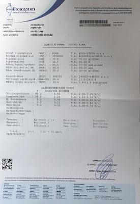
January 3rd, 2019
Blood tests
Blood tests (January 3rd, 2019) following the end of the first 3 months cycle of chemotherapy
Carbohydrate antigen 19-9 (CA 19-9): 273


===================================================
The patient started in January 21, 2019 a new 3 months cycle of chemotherapy with
FOLFIRINOX (modified)
Campto (Irinotecam), Eloxatin (Oxaliplatin), Leucovorin, 5FU
Η ασθενής άρχισε στις 21/01/2019 νέο κύκλο τρίμηνης χημειοθεραπείας με
FOLFIRINOX (modified)
Campto (Irinotecam), Eloxatin (Oxaliplatin), Leucovorin, 5FU
===================================================
April 19, 2019
UPPER AND LOWER ABDOMEN CT
Upper and Lower and Chest CT of April 19, 2019, following the end of the second 3 months cycle of chemotherapy
April 19, 2019
UPPER AND LOWER ABDOMEN CT
CT RIGHT HIP CT
TECHNIQUE The area of the upper and lower abdomen was covered with multisection highresolution CT technique with intravenous injection of contrast agent. Then transversal reconstructions were made of 7 mm thickness. Non-radiopaque medium per os administered. Fine transversal sections were obtained of 5 mm thickness at the level of the right hip and then 2-D coronal reconstructions were performed.
FINDINGS
Compared to prior upper lower abdomen CT at 07/01/2019. Diffused hepatic steatosis. In the middle of it, focal lesions are discernible with hypodense center and peripheral contrast enhancement, with smaller sizes in total versus prior one. Indicatively, lesion at VIII segment (line 3, image 418/1081) has at present maximal diameter 11 mm versus 18 mm at prior one, lesion at III segment (image 478) 21 mm versus 42 mm at prior one. To be noted that some of the preexisting foci have lost the hypodense center thereof and are mostly exhibited only as areas of increased contrast enhancement, such as a lesion at IV segment (image 515). Similar change presents also a preexisting lesion at the posterior crest of VII segment (image 522). Some smaller focal lesions at prior test are indiscernible at present. Significant reduction of the size exhibits also the preexisting primary space-occupying lesion with focus thereof the uncinate process of pancreas, at present test of maximal transversal size 41 x 24 mm (image 547) versus 55 x 36 mm at prior one. Is not exhibiting central necrosis at present test. It still obstructs the upper mesenteric vein and infiltrates at radius 360° the upper mesenteric artery. Small dotted air bubble is depicted at the right crest of the lesion, could be associated with infiltration of the duodenum. Pancreatic duct or the biliary tree without dilation. Collateral venal network at mesentery and omentum, in the context of the preexisting venal obstruction. Small nodular focus is depicted again at the body of the right adrenal, most likely related to adenoma. Left adrenal, spleen and kidneys normal. Small pleural effusion at pelvis minor, mildly increased versus prior test. Signs of turbidity depicted at the mesenteric root and marginally enlarged lymph nodes respectively, which however at present test exhibit mostly confluent morphology, directly below the lesion (image 601). The internal genitalia are without pathologic lesions.
-2nd page follows-
Intense lumbar spine scoliosis with curvature to the left and degenerative lesions. No focal lesions noted suspicious of secondary localizations at bone structures. Mild degenerative signs at the right hip joint, without image of fracture or suspicious focal lesions.
CONCLUSION
Reduction per size of the primary lesion at pancreas, as well as of secondary localizations at liver. Small increase of the pleural effusion volume at pelvis minor. Slightly increased size and confluent morphology of the pre-existing marginal lymph nodes at the mesenteric root.
CHEST CT
TECHNIQUE
The area of the chest was covered with multisection high-resolution CT technique of 0.6 mm thickness. Then reconstructions were made of 1.5 mm and 7 mm thickness as well as transversal MIP reconstructions. Contrast agent intravenously administered.
FINDINGS
Compared to prior chest CT from 07/01/2019. Without signs of secondary localizations or active pathology of another nature. Mild thick pleural – fibrotic signs at pulmonary apexes. Small extend atelectatic signs at the underlying surface of both the pulmonary bases, as well as small atelectatic bronchiectases at the posterior basal segment of the lower right lobe, within thereof dotted secretions are discernible. Without pleural or pericardial fluid effusion. Without enlarged lymph nodes at mediastinum, pulmonary portals and axillary cavities. Preexisting slightly projecting right paratracheal lymph nodes not differentiated. Mild calcifications of the coronal vessels and mitral valve. Size of the cardiovascular formation within the normal range. Without suspicious bone foci.
CONCLUSION
Without findings suspicious of secondary localizations and without changes versus prior imaging test (1st/2019).
Download and Review the CT
Κατεβάστε και Δείτε την αξονική τομογραφία
One Drive page
CT scan 19-4-2019
Left click on the above file, then right click on the name of the folder (CT scan 7-01-2018) and then
click on "download" to obtain the zip file (wait for some minutes). Open this file by double left clicking on it and then double left click
on ''startcd.exe'' file and then click on "extract files" to have all the cd files. Double click then on startcd.exe file and then on "open the viewer" to start reviewing the CT
Πατείστε επάνω στις εικόνες για να μεγαλώσουν


===================================================
April 15, 2019
Blood tests
Blood tests (April 15, 2019) following the end of the first 3 months cycle of chemotherapy
Carbohydrate antigen 19-9 (CA 19-9): 44,8


===================================================
The patient is taken the same therapy for another 3-months cycle of 6 treatments
Η ασθενής συνεχίζει την ίδια θεραπεία για άλλο ένα κύκλο 3 μηνών με 6 αγωγές
An interesting fact is that after the second treatment both the cancer markers are normal, as:
CA 19-9 : 18,6 U/mL and CEA : 4,6 ng/mL
Μια ενδιαφέρουσα παρατήρηση είναι ότι ήδη μετά την δεύτερη αγωγή και οι 2 καρκινικοί δείκτες είναι φυσιολογικοί, ήτοι
CA 19-9 : 18,6 U/mL and CEA : 4,6 ng/mL
===================================================
August 23, 2019
UPPER AND LOWER ABDOMEN CT
Upper and Lower and Chest CT of August 19, 2019, following the end of the third 3 months cycle of chemotherapy
August 23, 2019
UPPER & LOWER ABDOMEN CT
TECHNIQUE The area of the upper and lower abdomen was covered with multislice high-resolution CT technique with intravenous contrast agent injection. Then, transversal reconstructions were made of 7 mm thickness. Non-radiopaque per os medium administered.
FINDINGS Study compared to prior upper-lower abdomen CT from 19-04-2019. The diffused hepatic steatosis is depicted again. Within thereof focal lesions are noted with hypodense center and peripheral contrast enhancement. Lesion at the VIII segment (image 349/873) has maximal diameter about 11 mm as in prior study while lesion at III segment (image 375/873) has at present maximal diameter 13 mm versus 20 mm at prior study. Lesion at the IV segment (image 430) is depicted again without major differentiation as area of increased contrast enhancement. To be noted that at present study prior existing lesions depicted as areas of increased enhancement at the posterior crest of the VII segment are depicted today with hypodense center and have maximal size 13 mm and 11 mm respectively (image 457/873). The prior existing primary space-occupying lesion is depicted again focusing on the uncinate process of the pancreas with minor degree increase of the size thereof and maximal transversal size at present test 47 x 26 mm versus 41 x 24 mm alongside prior study while novel finding consists a small area of central necrosis. It continues to obstruct the upper mesenteric vein and infiltrate at a radius of 360° the upper mesenteric artery which is made indiscernible at small extend with presence however of more peripheral flow. The possibility of duodenal infiltration cannot be excluded. No dilation of the biliary tree or pancreatic duct noted. The collateral venal network is depicted again at mesentery and omentum in the context of the prior mentioned venal obstruction. Small nodular focus is depicted again at the body of the right adrenal, most likely of adenomatous origin without significant differentiation. Spleen, left adrenal and kidneys are depicted normally. No signs of obstructive uropathy noted. The signs of turbidity at mesenteric root are depicted again as well as the confluent mesenteric lymph nodes at said level, below the lesion without major differentiation. The prior existing moderate pleural fluid effusion is depicted again at pelvis minor. Minor degree mural lesion-edematous image of some of the small intestine helices at the area of pelvis minor (consists novel finding). Without pathological lesions from the internal genitalia. Intense left tilted scoliosis of the lumbar spine and degenerative lesions.
Degenerative signs depicted also at the right hip joint. Novel finding consists the upper epiphyseal surface sclerosis and reduction of the height of the L4 vertebra’s body. Upon clinical indications more specific imaging study is recommended.
CONCLUSION
Minor degree increase of size of the primary lesion at pancreas which infiltrates the upper mesenteric artery which is made indiscernible at minor extend (novel finding). Minor degree reduction of the secondary localization at liver while another one is depicted without notable differentiation. Two other secondary localizations are depicted with differentiation of their morphology presenting hypodense center at present study versus areas of increased contrast enhancement at prior study. The pleural fluid effusion at pelvis minor is not depicted at prior study while novel finding consist the few small intestine helices with thickened wall. Without notable differentiation of the pre-existing marginal lymph nodes at the mesenteric root with confluent morphology. Rest of the findings as described above.
CHEST CT
TECHNIQUE The area of the chest was covered with multislice high-resolution CT technique of 0.6 mm thickness. Then reconstructions were made of 1.5 mm and 7 mm thickness as well as transversal MIP reconstructions. Contrast agent intravenously injected.
FINDINGS Study compared to prior chest CT from 19/04/2019. The mild thick pleural-fibrotic signs at both the pulmonary apexes are depicted again. No findings suspicious of secondary localizations noted from the study of pulmonary parenchyma. The small atelectatic bronchiectases at the posterior basal segment of the lower right lobe are depicted again with dotted excretions within thereof. Novel finding consists the very small pleural fluid effusion at left as well as the small extend atelectatic signs at the left pulmonary base. Segmental atelectasis of the apical segment of the lower left lobe consists also novel finding. Without pericardial or pleural fluid effusion at right. No pathologically enlarged lymph nodes depicted at mediastinum, pulmonary portals and axillary areas. Pre-existing slightly projecting right paratracheal lymph nodes not presenting major differentiation while a few lymph nodes by the aortic arch are more visible at prior study with size however within the normal range. Mild calcifications at coronal vessels and mitral valve as well as presence of calcifications at the thoracic aorta. The cardiovascular formation is depicted with normal size. Degenerative lesions at the thoracic spine without further findings. Absence of contrast enhancement at the left brachiocephalic vein alongside the catheter’s course, a finding suspicious of thrombosis, further study with computed venography is recommended.
CONCLUSION
Findings suspicious of secondary localizations not detected also at present study. Novel finding consists the small pleural fluid effusion at left as well as the atelectatic sings at the left pulmonary base and at the apical segment of the lower left lobe. Novel finding consists also the absence of contrast enhancement at the left brachiocephalic vein alongside the catheter’s course, a finding suspicious of thrombosis, further study with computed venography is recommended. Rest of the findings as described above.
Αξονική Τομογραφία της 23ης Αυγούστου 2019, μετά το πέρας
του δεύτερου τρίμηνου κύκλου της χημειοθεραπείας
FOLFIRINOX


===================================================
Μετά τη διαπίστωση της θρόμβωσης και πλήρους απόφραξης της βραχιονοκεφαλικής φλέβας εντός της οποίας είχε τοποθετηθεί ο καθετήρας του portacath, έγινε η αφαίρεσή του και η τοποθέτηση νέου portacath στην δεξιά πλευρά του στήθους, με τον καθετήρα εντός της σφαγίτιδας φλέβας.
Εν συνεχεία απεφασίσθη η συνέχιση της θεραπείας με FOLFIRINOX για 1 εισέτι τρίμηνο με αρχή γενομένη την 30τήν Σεπτεμβρίου 2019
===================================================
For more, contact
Marios Leftheriotis
e-mail
marleft@otenet.gr
click on "download" to obtain the zip file (wait for some minutes). Open this file by double left clicking on it and then double left click
on ''startcd.exe'' file and then click on "extract files" to have all the cd files. Double click then on startcd.exe file and then on "open the viewer" to start reviewing the CT
or
Download in DROPBOX
https://www.dropbox.com/s/ti86p5szpzclbjj/CT%20scan%207-01-2019%20ISO%20file.zip?dl=0
Πατείστε επάνω στις εικόνες για να μεγαλώσουν


===================================================
January 3rd, 2019
Blood tests
Blood tests (January 3rd, 2019) following the end of the first 3 months cycle of chemotherapy
Carbohydrate antigen 19-9 (CA 19-9): 273
January 3rd, 2019
Blood tests
Click on the following
texts to enlarge them


===================================================
The patient started in January 21, 2019 a new 3 months cycle of chemotherapy with
FOLFIRINOX (modified)
Campto (Irinotecam), Eloxatin (Oxaliplatin), Leucovorin, 5FU
Η ασθενής άρχισε στις 21/01/2019 νέο κύκλο τρίμηνης χημειοθεραπείας με
FOLFIRINOX (modified)
Campto (Irinotecam), Eloxatin (Oxaliplatin), Leucovorin, 5FU
===================================================
April 19, 2019
UPPER AND LOWER ABDOMEN CT
Upper and Lower and Chest CT of April 19, 2019, following the end of the second 3 months cycle of chemotherapy
Name M.L.
Age 77
UPPER AND LOWER ABDOMEN CT
CT RIGHT HIP CT
TECHNIQUE The area of the upper and lower abdomen was covered with multisection highresolution CT technique with intravenous injection of contrast agent. Then transversal reconstructions were made of 7 mm thickness. Non-radiopaque medium per os administered. Fine transversal sections were obtained of 5 mm thickness at the level of the right hip and then 2-D coronal reconstructions were performed.
FINDINGS
Compared to prior upper lower abdomen CT at 07/01/2019. Diffused hepatic steatosis. In the middle of it, focal lesions are discernible with hypodense center and peripheral contrast enhancement, with smaller sizes in total versus prior one. Indicatively, lesion at VIII segment (line 3, image 418/1081) has at present maximal diameter 11 mm versus 18 mm at prior one, lesion at III segment (image 478) 21 mm versus 42 mm at prior one. To be noted that some of the preexisting foci have lost the hypodense center thereof and are mostly exhibited only as areas of increased contrast enhancement, such as a lesion at IV segment (image 515). Similar change presents also a preexisting lesion at the posterior crest of VII segment (image 522). Some smaller focal lesions at prior test are indiscernible at present. Significant reduction of the size exhibits also the preexisting primary space-occupying lesion with focus thereof the uncinate process of pancreas, at present test of maximal transversal size 41 x 24 mm (image 547) versus 55 x 36 mm at prior one. Is not exhibiting central necrosis at present test. It still obstructs the upper mesenteric vein and infiltrates at radius 360° the upper mesenteric artery. Small dotted air bubble is depicted at the right crest of the lesion, could be associated with infiltration of the duodenum. Pancreatic duct or the biliary tree without dilation. Collateral venal network at mesentery and omentum, in the context of the preexisting venal obstruction. Small nodular focus is depicted again at the body of the right adrenal, most likely related to adenoma. Left adrenal, spleen and kidneys normal. Small pleural effusion at pelvis minor, mildly increased versus prior test. Signs of turbidity depicted at the mesenteric root and marginally enlarged lymph nodes respectively, which however at present test exhibit mostly confluent morphology, directly below the lesion (image 601). The internal genitalia are without pathologic lesions.
-2nd page follows-
Intense lumbar spine scoliosis with curvature to the left and degenerative lesions. No focal lesions noted suspicious of secondary localizations at bone structures. Mild degenerative signs at the right hip joint, without image of fracture or suspicious focal lesions.
CONCLUSION
Reduction per size of the primary lesion at pancreas, as well as of secondary localizations at liver. Small increase of the pleural effusion volume at pelvis minor. Slightly increased size and confluent morphology of the pre-existing marginal lymph nodes at the mesenteric root.
CHEST CT
TECHNIQUE
The area of the chest was covered with multisection high-resolution CT technique of 0.6 mm thickness. Then reconstructions were made of 1.5 mm and 7 mm thickness as well as transversal MIP reconstructions. Contrast agent intravenously administered.
FINDINGS
Compared to prior chest CT from 07/01/2019. Without signs of secondary localizations or active pathology of another nature. Mild thick pleural – fibrotic signs at pulmonary apexes. Small extend atelectatic signs at the underlying surface of both the pulmonary bases, as well as small atelectatic bronchiectases at the posterior basal segment of the lower right lobe, within thereof dotted secretions are discernible. Without pleural or pericardial fluid effusion. Without enlarged lymph nodes at mediastinum, pulmonary portals and axillary cavities. Preexisting slightly projecting right paratracheal lymph nodes not differentiated. Mild calcifications of the coronal vessels and mitral valve. Size of the cardiovascular formation within the normal range. Without suspicious bone foci.
CONCLUSION
Without findings suspicious of secondary localizations and without changes versus prior imaging test (1st/2019).
Download and Review the CT
Κατεβάστε και Δείτε την αξονική τομογραφία
One Drive page
CT scan 19-4-2019
click on "download" to obtain the zip file (wait for some minutes). Open this file by double left clicking on it and then double left click
on ''startcd.exe'' file and then click on "extract files" to have all the cd files. Double click then on startcd.exe file and then on "open the viewer" to start reviewing the CT
Πατείστε επάνω στις εικόνες για να μεγαλώσουν


===================================================
April 15, 2019
Blood tests
Blood tests (April 15, 2019) following the end of the first 3 months cycle of chemotherapy
Carbohydrate antigen 19-9 (CA 19-9): 44,8
April 15, 2019
Blood tests
Click on the following
texts to enlarge them


===================================================
The patient is taken the same therapy for another 3-months cycle of 6 treatments
Η ασθενής συνεχίζει την ίδια θεραπεία για άλλο ένα κύκλο 3 μηνών με 6 αγωγές
An interesting fact is that after the second treatment both the cancer markers are normal, as:
CA 19-9 : 18,6 U/mL and CEA : 4,6 ng/mL
Μια ενδιαφέρουσα παρατήρηση είναι ότι ήδη μετά την δεύτερη αγωγή και οι 2 καρκινικοί δείκτες είναι φυσιολογικοί, ήτοι
CA 19-9 : 18,6 U/mL and CEA : 4,6 ng/mL
===================================================
August 23, 2019
UPPER AND LOWER ABDOMEN CT
Upper and Lower and Chest CT of August 19, 2019, following the end of the third 3 months cycle of chemotherapy
Name M.L.
Age 78
UPPER & LOWER ABDOMEN CT
TECHNIQUE The area of the upper and lower abdomen was covered with multislice high-resolution CT technique with intravenous contrast agent injection. Then, transversal reconstructions were made of 7 mm thickness. Non-radiopaque per os medium administered.
FINDINGS Study compared to prior upper-lower abdomen CT from 19-04-2019. The diffused hepatic steatosis is depicted again. Within thereof focal lesions are noted with hypodense center and peripheral contrast enhancement. Lesion at the VIII segment (image 349/873) has maximal diameter about 11 mm as in prior study while lesion at III segment (image 375/873) has at present maximal diameter 13 mm versus 20 mm at prior study. Lesion at the IV segment (image 430) is depicted again without major differentiation as area of increased contrast enhancement. To be noted that at present study prior existing lesions depicted as areas of increased enhancement at the posterior crest of the VII segment are depicted today with hypodense center and have maximal size 13 mm and 11 mm respectively (image 457/873). The prior existing primary space-occupying lesion is depicted again focusing on the uncinate process of the pancreas with minor degree increase of the size thereof and maximal transversal size at present test 47 x 26 mm versus 41 x 24 mm alongside prior study while novel finding consists a small area of central necrosis. It continues to obstruct the upper mesenteric vein and infiltrate at a radius of 360° the upper mesenteric artery which is made indiscernible at small extend with presence however of more peripheral flow. The possibility of duodenal infiltration cannot be excluded. No dilation of the biliary tree or pancreatic duct noted. The collateral venal network is depicted again at mesentery and omentum in the context of the prior mentioned venal obstruction. Small nodular focus is depicted again at the body of the right adrenal, most likely of adenomatous origin without significant differentiation. Spleen, left adrenal and kidneys are depicted normally. No signs of obstructive uropathy noted. The signs of turbidity at mesenteric root are depicted again as well as the confluent mesenteric lymph nodes at said level, below the lesion without major differentiation. The prior existing moderate pleural fluid effusion is depicted again at pelvis minor. Minor degree mural lesion-edematous image of some of the small intestine helices at the area of pelvis minor (consists novel finding). Without pathological lesions from the internal genitalia. Intense left tilted scoliosis of the lumbar spine and degenerative lesions.
Degenerative signs depicted also at the right hip joint. Novel finding consists the upper epiphyseal surface sclerosis and reduction of the height of the L4 vertebra’s body. Upon clinical indications more specific imaging study is recommended.
CONCLUSION
Minor degree increase of size of the primary lesion at pancreas which infiltrates the upper mesenteric artery which is made indiscernible at minor extend (novel finding). Minor degree reduction of the secondary localization at liver while another one is depicted without notable differentiation. Two other secondary localizations are depicted with differentiation of their morphology presenting hypodense center at present study versus areas of increased contrast enhancement at prior study. The pleural fluid effusion at pelvis minor is not depicted at prior study while novel finding consist the few small intestine helices with thickened wall. Without notable differentiation of the pre-existing marginal lymph nodes at the mesenteric root with confluent morphology. Rest of the findings as described above.
CHEST CT
TECHNIQUE The area of the chest was covered with multislice high-resolution CT technique of 0.6 mm thickness. Then reconstructions were made of 1.5 mm and 7 mm thickness as well as transversal MIP reconstructions. Contrast agent intravenously injected.
FINDINGS Study compared to prior chest CT from 19/04/2019. The mild thick pleural-fibrotic signs at both the pulmonary apexes are depicted again. No findings suspicious of secondary localizations noted from the study of pulmonary parenchyma. The small atelectatic bronchiectases at the posterior basal segment of the lower right lobe are depicted again with dotted excretions within thereof. Novel finding consists the very small pleural fluid effusion at left as well as the small extend atelectatic signs at the left pulmonary base. Segmental atelectasis of the apical segment of the lower left lobe consists also novel finding. Without pericardial or pleural fluid effusion at right. No pathologically enlarged lymph nodes depicted at mediastinum, pulmonary portals and axillary areas. Pre-existing slightly projecting right paratracheal lymph nodes not presenting major differentiation while a few lymph nodes by the aortic arch are more visible at prior study with size however within the normal range. Mild calcifications at coronal vessels and mitral valve as well as presence of calcifications at the thoracic aorta. The cardiovascular formation is depicted with normal size. Degenerative lesions at the thoracic spine without further findings. Absence of contrast enhancement at the left brachiocephalic vein alongside the catheter’s course, a finding suspicious of thrombosis, further study with computed venography is recommended.
CONCLUSION
Findings suspicious of secondary localizations not detected also at present study. Novel finding consists the small pleural fluid effusion at left as well as the atelectatic sings at the left pulmonary base and at the apical segment of the lower left lobe. Novel finding consists also the absence of contrast enhancement at the left brachiocephalic vein alongside the catheter’s course, a finding suspicious of thrombosis, further study with computed venography is recommended. Rest of the findings as described above.
Αξονική Τομογραφία της 23ης Αυγούστου 2019, μετά το πέρας
του δεύτερου τρίμηνου κύκλου της χημειοθεραπείας
FOLFIRINOX


COMPUTED VENOGRAPHY
of the brachiocephalic vein to verify thrombosis and complete obstruction of this vein
Conclusion
Presence of portacath in left brachiocephalic vein with
thrombosis and occlusion
ΑΞΟΝΙΚΗ ΦΛΕΒΟΓΡΑΦΙΑ ΑΡΙΣΤΕΡΟΥ ΑΝΩ ΑΚΡΟΥ
της 9ης Σεπτεμβρίου 2019, προς διαπίστωση της θρόμβωσης και πλήρους απόφραξης της βραχιονοκεφαλικης φλέβας εντός
της οποίας ήταν τοποθετημένος ο καθετήρας του portacath

===================================================
After thrombosis and complete obstruction of the brachiocephalic vein into which the portacath catheter was inserted, the catheter was removed
and a new portacath was placed on the right side of the chest, with the catheter
inserted into the jugular vein.
Subsequently, it was
decided to continue treatment with FOLFIRINOX for a period of 3 months starting on September 30, 2019.
Μετά τη διαπίστωση της θρόμβωσης και πλήρους απόφραξης της βραχιονοκεφαλικής φλέβας εντός της οποίας είχε τοποθετηθεί ο καθετήρας του portacath, έγινε η αφαίρεσή του και η τοποθέτηση νέου portacath στην δεξιά πλευρά του στήθους, με τον καθετήρα εντός της σφαγίτιδας φλέβας.
Εν συνεχεία απεφασίσθη η συνέχιση της θεραπείας με FOLFIRINOX για 1 εισέτι τρίμηνο με αρχή γενομένη την 30τήν Σεπτεμβρίου 2019
===================================================
For more, contact
Marios Leftheriotis
marleft@otenet.gr
Personal Page